


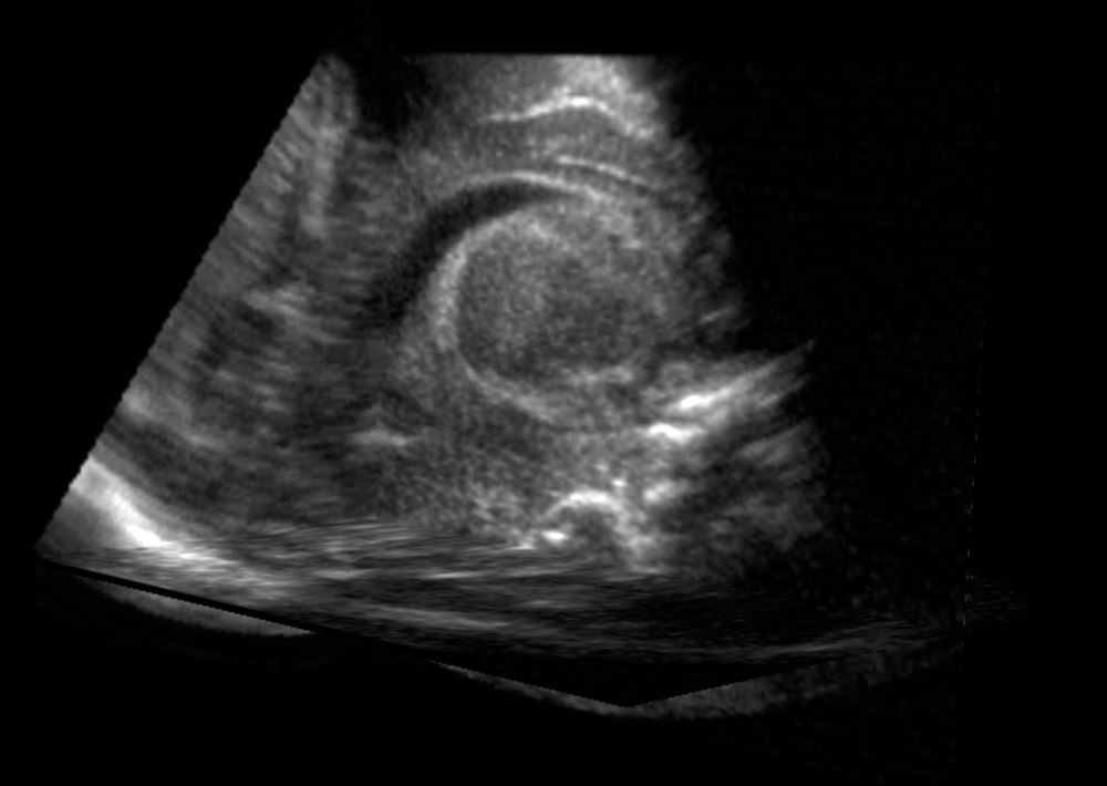
Pregnant women across the country walk into clinics expecting reassurance. They leave with printed images of a fetus and a feeling that everything is being monitored, measured, and managed. The ultrasound has become a ritual of modern pregnancy. It is marketed as safe, routine, and emotionally meaningful.
What it is not marketed as is what it actually is: a pulse of high-intensity acoustic radiation directed at one of the most biologically sensitive objects in human development.
This is not a fringe position. The physics of ultrasound are not in dispute. The biological mechanisms by which acoustic energy interacts with living tissue are not in dispute. What is in dispute is whether the medical industry has done the work necessary to justify the current frequency of prenatal scans, and whether the public has been given accurate information to make that judgment for themselves.
The answer to both questions is no.
THE MECHANICS OF WHAT'S ACTUALLY HAPPENING
Ultrasound is not a camera. The comparison is wrong and it matters that it's wrong.
A camera captures light that is already present. It is passive. It records. Ultrasound works by generating and transmitting energy into tissue. It is active. It fires.
The technology uses high-frequency sound waves, typically between 2 and 18 megahertz for diagnostic purposes, pulsed into the body from a transducer. Those waves travel through tissue, reflect off structures with different acoustic densities, and return to the transducer where the machine assembles them into an image. The image is a byproduct. The waves are the mechanism.
When those waves move through tissue, they do not pass through without consequence. Acoustic energy interacts with the cellular environment in ways that produce measurable physical effects. The two most significant are thermal effects and mechanical effects.
Thermal effects are straightforward: acoustic energy converts to heat in tissue. The degree of heating depends on the intensity of the beam, the duration of exposure, and the acoustic absorption properties of the tissue being scanned. Bone absorbs more acoustic energy than soft tissue and heats faster. The fetal skull, the developing vertebrae, and the calcifying structures of a growing skeleton are particularly susceptible. Regulatory guidelines set limits on thermal index values, but those limits are based on models, not on comprehensive studies of developmental outcome in humans.
Mechanical effects are where it gets more complicated. The most significant mechanical effect is acoustic cavitation.
Cavitation occurs when the alternating pressure of sound waves causes dissolved gases in biological fluid to form bubbles. These bubbles oscillate with the pressure cycles. Under stable conditions, this oscillation is called stable cavitation. Under higher intensities, the bubbles grow rapidly and then collapse violently. This is called inertial or transient cavitation.
The collapse of cavitation bubbles is not a minor event at the cellular scale. The implosion generates localized heat spikes, mechanical shockwaves, and free radical production. In adult tissue, the effects are transient and the body's repair mechanisms manage them. In a developing fetus, particularly in the first and second trimesters, the cellular environment is fundamentally different. Tissues are forming, not maintaining. Neurons are migrating, not stabilizing. The biological resilience that adult tissue has is not yet present.
This is not a theoretical concern invented by critics of the medical industry. It is a known property of acoustic physics that was documented before prenatal ultrasound became a routine procedure. The question of whether routine diagnostic ultrasound produces cavitation in fetal tissue at standard clinical settings remains genuinely unresolved. The FDA and the industry that manufactures ultrasound equipment acknowledge this uncertainty while simultaneously expanding the technology and increasing its use.
That is the core problem.
WHAT THE YALE RESEARCH SHOWED
In 2006, a research team led by Dr. Pasko Rakic at Yale University published findings in the in the Proceedings of the National Academy of Sciences that deserve significantly more attention than they received. The study examined the effect of prenatal ultrasound exposure on neuronal migration in fetal mice.
Neuronal migration is the process by which neurons produced in the inner layers of the developing brain travel outward to their designated positions in the cortex. This process follows a precise spatial and temporal pattern. Neurons are produced in sequence, and they migrate in sequence, forming the layered architecture of the cortex in a specific order. Disruption of this process does not produce a different brain. It produces a disorganized one.
What Rakic's team found was that mouse fetuses exposed to ultrasound showed statistically significant disruption of neuronal migration. A measurable percentage of neurons failed to reach their proper cortical positions. They remained displaced, distributed through layers where they did not belong.
The study was carefully designed. It used ultrasound equipment comparable to clinical diagnostic devices. It controlled for exposure duration and timing within the gestational period. It used markers to track individual neurons and confirm their final positions. The findings were not ambiguous.
Rakic himself was careful in how he framed the results. He noted that the mouse brain develops on a different timeline than the human brain, and that direct translation of mouse findings to human clinical practice requires caution. He called for further study. He was not calling for a ban. He was flagging a signal that required investigation.
That investigation, at the scale and rigor that the finding warranted, did not happen. The ultrasound industry continued to expand. The number of scans per pregnancy continued to increase. High-definition and 3D ultrasound technology, which operates at higher intensities and longer exposure durations, became commercially available and was marketed directly to pregnant women as an enhancement of the bonding experience.
The signal from the 2006 Yale research was not refuted. It was absorbed into the background noise of an industry that had no financial incentive to pursue it seriously.
THE NEURODEVELOPMENTAL CORRELATION
The United States has seen a documented and substantial increase in neurodevelopmental disorders over the past four decades. Autism spectrum disorder diagnoses have increased by orders of magnitude. Attention deficit disorders, sensory processing disorders, and speech and language delays have all increased in prevalence during the same period.
Identifying causation in complex biological outcomes is difficult. Multiple environmental factors have changed simultaneously. Diagnostic criteria have expanded. Awareness has increased. These variables are real and they complicate any single-factor analysis.
None of that changes the following fact: the period during which prenatal ultrasound moved from limited medical use to routine and frequent application overlaps precisely with the period during which neurodevelopmental disorder rates began their sustained increase.
This is a correlation. Correlation does not establish causation. That is a true statement. It is also a statement that is frequently used to shut down inquiry rather than advance it. The appropriate response to a correlation between a widespread prenatal exposure and a documented increase in neurodevelopmental outcomes is rigorous investigation. What has not occurred is rigorous investigation funded by the entities that profit from that exposure.
The studies that exist on prenatal ultrasound safety are largely short-term, focused on immediate adverse events rather than long-term developmental outcomes, and many are funded or conducted by parties with financial relationships to the ultrasound industry. This is not a conspiracy. It is a standard feature of how medical research gets funded in the United States. Industries fund research into their products. Funding shapes questions. Questions shape findings. The absence of evidence is not the same as evidence of absence, and in the context of prenatal ultrasound safety, the absence of long-term, independently funded, longitudinal studies of neurological outcomes is a critical gap.
Epidemiological data from the United States does not stand alone. In countries where routine prenatal ultrasound was adopted later or used less frequently, the comparable explosion in neurodevelopmental disorder rates either lagged or was less pronounced. This observation does not settle the question. It keeps the question open in a way that honest science requires.
THE FREQUENCY PROBLEM
In the 1970s, when prenatal ultrasound was introduced into clinical practice in the United States, a typical pregnancy might involve one or two scans. The clinical justification was specific: confirm viability, check placental position, assess fetal growth when clinical concerns existed.
Today the number of scans per pregnancy has increased substantially. Some practices recommend first-trimester dating scans, nuchal translucency screening scans at 11 to 13 weeks, anatomy scans at 18 to 20 weeks, growth scans in the third trimester, and additional scans whenever a question arises. Women carrying multiples, women with gestational diabetes, women classified as high-risk for any reason, may receive a scan at nearly every prenatal visit.
Beyond the clinical setting, the ultrasound industry developed and aggressively marketed keepsake or boutique ultrasound services. These are commercial operations that sell extended 3D and 4D imaging sessions to pregnant women who want longer views of their developing child. These sessions involve no clinical justification. They are purely commercial. They use higher-resolution technology that requires more intense acoustic output to produce the cleaner images that customers are paying for. The FDA has issued statements discouraging elective keepsake ultrasounds. Those statements exist as text on a government website. They have had no meaningful effect on the market.
The cumulative acoustic exposure of a fetus in a pregnancy with frequent clinical scans plus one or more commercial sessions is substantially different from the exposure associated with the limited diagnostic use for which the technology's safety profile was originally assessed. The industry does not discuss this distinction in its public communications. Prenatal care providers, with notable exceptions, do not raise it with patients. The default assumption is that more information is better and that the cost of obtaining that information is zero.
That assumption has not been validated. It was adopted because it served the interests of parties who profit from it, and it was maintained because no powerful institutional voice had reason to challenge it.
HOW SAFETY GUIDELINES ACTUALLY WORK
The FDA regulates diagnostic ultrasound devices. The current regulatory framework uses two indices to limit potential biological effects during clinical use: the thermal index, which estimates the potential for tissue heating, and the mechanical index, which estimates the potential for cavitation effects.
These indices are displayed on ultrasound machines in real time. The FDA sets upper limits. Clinical guidelines from professional obstetric organizations recommend keeping exposure to the lowest levels consistent with obtaining diagnostically useful information. This principle is called ALARA: as low as reasonably achievable.
The ALARA principle is sound. Its implementation is inconsistent. The operators of ultrasound machines, including sonographers and physicians, set the machine parameters. Training in acoustic output management varies. In a busy clinical environment, the pressure to obtain clear, useful images quickly does not always align with the discipline of minimizing output levels. Commercial keepsake operations have no clinical obligation to follow ALARA at all.
The thermal and mechanical indices are models. They are calculated estimates based on assumptions about tissue properties and acoustic behavior. They are not direct measurements of what is happening in the specific tissue being imaged. For fetal brain tissue at various stages of development, the models carry significant uncertainty. Fetal brain tissue has different acoustic properties than adult brain tissue. The models were validated primarily on adult tissue data.
The safety guidelines were constructed by organizations with institutional relationships to the medical device industry. The FDA device approval process for ultrasound equipment does not require manufacturers to demonstrate safety through long-term developmental outcome data. It requires manufacturers to demonstrate that devices operate within defined output parameters. Whether those parameters are the right parameters, based on the right biological models, assessed against the right outcome measures, is a question the approval process does not answer.
THE REVENUE STRUCTURE
Prenatal care in the United States generates substantial revenue. Ultrasound scanning is one of the most frequently performed and billed procedures in obstetrics. The equipment costs are high but the per-procedure costs are relatively low once equipment is purchased. The margin structure strongly incentivizes high scan volume.
The manufacturers of ultrasound equipment are large medical device companies with significant lobbying presence and research relationships with academic medical centers. The professional organizations that set clinical guidelines for obstetric care include members with financial ties to those manufacturers. This does not mean that every guideline is corrupt. It means that the institutional structure through which safety questions would need to be raised and investigated is occupied by parties who have financial reasons not to raise them aggressively.
Independent researchers who raise safety concerns about prenatal ultrasound face predictable institutional friction. Publication in high-impact journals requires peer review by researchers in the field, many of whom have professional and financial relationships with the technology. Funding for follow-up research on safety signals depends on grant mechanisms that are shaped by institutional priorities. An academic researcher who builds a career around questioning prenatal ultrasound safety is operating against the grain of the institutional incentives that govern how medical research careers are built.
This is not unique to ultrasound. It is a general feature of how safety questions get handled when the technology in question is profitable and already deeply embedded in clinical practice. The same structural dynamics appeared in the histories of other medical technologies that were eventually found to carry risks that were not initially acknowledged.
WHAT ADEQUATE RESEARCH WOULD LOOK LIKE
A properly designed long-term safety study of prenatal ultrasound would require several components that no such study has yet combined.
It would track a large cohort of children from birth, with precise documentation of prenatal ultrasound exposure including number of scans, gestational age at each scan, scan duration, and machine output settings. It would follow those children through at minimum school age, with standardized neurodevelopmental assessments at multiple time points. It would be funded by sources without financial interest in the outcome. It would be designed and conducted by researchers without financial relationships to the ultrasound industry. It would have preregistered endpoints to prevent the post-hoc redefinition of outcomes that allows studies to report negative findings when positive ones were actually present.
This study does not exist. The studies that do exist are shorter, smaller, less precisely documented in terms of actual exposure, and conducted under institutional conditions that do not favor finding adverse effects. When researchers cite the existing literature to conclude that prenatal ultrasound is safe, they are accurately representing what the existing literature shows. They are not accurately representing the adequacy of that literature to answer the question it is being cited to answer.
The absence of a properly designed safety study is not a technical problem. Conducting such a study is feasible. The infrastructure exists. The cohort sizes are achievable. The cost is within the range of research that gets funded regularly for questions of comparable public health significance. The reason the study has not been conducted is that the parties with the resources and institutional standing to conduct it have not chosen to do so. The parties who have raised the need for it do not have those resources.
WHAT YOU SHOULD KNOW BEFORE YOUR NEXT SCAN
None of this is an argument that prenatal ultrasound has no value. It does have value. Identifying ectopic pregnancies, assessing fetal position, evaluating placenta previa, measuring growth in high-risk pregnancies: these are legitimate clinical applications where the information obtained addresses a specific clinical question and the benefit of that information is concrete.
The argument is different from that. The argument is that the number of scans in a typical low-risk pregnancy in the United States today exceeds what the clinical evidence justifies, that the safety profile of that level of exposure has not been adequately studied, and that the system through which that question would be investigated and answered is structurally compromised by the financial interests of the parties who would need to investigate it.
If you are pregnant, or planning to be, the following is worth knowing.
Every scan carries acoustic exposure. The cumulative total matters. A low-risk pregnancy that proceeds normally does not require a scan at every prenatal visit. The ALARA principle exists for a reason; asking your provider about it is a reasonable and appropriate thing to do.
The anatomy scan at 18 to 20 weeks and a limited first-trimester scan to confirm viability and dating represent a baseline of clinical utility that is well-supported. Scans beyond that in a low-risk pregnancy carry marginal additional clinical value and non-zero additional exposure.
Keepsake ultrasound sessions serve no clinical function. The information obtained from the Rakic research and from the acoustic physics literature is sufficient reason to decline them.
You are allowed to ask questions. You are allowed to request the minimum clinically necessary scan duration. You are allowed to decline scans that are offered as routine when your clinical situation does not make them necessary. These are not hostile positions. They are the exercise of informed consent, which is a legal and ethical right, and which requires that you actually be informed.
The information your care provider presents to you about prenatal ultrasound safety is shaped by guidelines from organizations with financial ties to the industry that produces the technology. That does not make your provider dishonest. It makes the information incomplete.
WHERE THIS LEAVES YOU
The developing brain operates on biological processes of extraordinary precision. Neurons move to specific locations at specific times according to signals that are not fully understood. The acoustic environment inside the uterus during a scan is substantially different from the acoustic environment in the absence of a scan. Whether that difference matters, at what exposure levels, during which windows of fetal development, is a question that has not been adequately answered.
The honest position is that the safety of routine frequent prenatal ultrasound, at the levels currently practiced in the United States, has not been established. It has been assumed. The assumption is profitable. The parties who benefit from it have not invested adequately in testing it. The regulatory framework that governs the technology was not designed to catch long-term developmental effects.
That is a different thing than safety. It is the appearance of safety, maintained by the absence of the research that would be required to establish it as fact.
You should know that. You should ask questions based on it. And you should make decisions about your pregnancy with that gap clearly in view, not obscured by the confidence of an industry that has not done the work to justify it.